Sunday, July 29, 2007

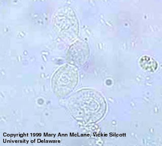
Pictures of urinary casts and other urinary cellular components


Spermatozoa


Renal Epithelial cells


Yeast cells (notice the budding)


Cellular cast


Granular cast


Hyaline cast


Sasi
Tg02
0503804g

Urinary Crystals pictures and brief infor (sasi's)


Uric Acid crystals


Calcium Oxalate crystal (look out for the 'X' )


Triple phosphate crystal (coffin like)

Crystal formation in urine can be caused:
- By an augmentation of concentration beyond the supersaturation capacity. This situation is mostly the result of a decreased dilution like in a case of insufficient water intake.
- By a decreased supersaturation capacity. This situation could be caused by a decrease in inhibitors concentration, a neutralization of these inhibitors, by some electrolytes, or a by pH change.
- By the presence of crystals with a promoter effect on the crystallization of another species. Crystallization of calcium oxalate promoted by amorphous urates is a good example of this phenomenon.

Usual urine crystals found in Urine:
Alkaline pH
Amorphous phosphates
Triple phosphates
Ammonium biurates
Calcium phosphates
Calcium carbonates
Acid pH
Amorphous urates
Uric acid
Calcium oxalates
Cystine
Sasi
Tg02
0503804g

Microbiology and Serology

Hi everyone,
Sasi here reporting from one of the private labs in Singapore. The first three weeks of my attachment I was attached to the Microbiology department. I basically had to run urine analysis via the URISYS 2400 urinalysis analyzer. The purpose of UAN (Urine Analysis) is for the semi-quantitative determination of pH, leukocytes, nitrites, protein, glucose, ketone bodies, urobilinogen, bilirubin, and erythrocytes in urine. Urine test strips are used to measure certain constituents in urine which signify renal, urinary, hepatic and metabolic disorders. In the mornings, I would start off by running the controls for the machine by loading the thawed controls. Then, I would record down both the positive and negative control results and make sure they tallied with the previous days' control results. In the afternoon, majority of the urine samples would begin to arrive. The urine specimen must be collected in a clean, dry container, either plastic or glass without preservatives. Once the specimens were received, they were labelled with the appropriate barcodes and loaded into the machine. When loading the samples, I had to make sure the sequence number on the monitor matched the first sample that was going to be loaded so that the results can be easily tracked down (via the sequence number) in the future in case of an emergency. This whole process of running the samples had to be done fast and accurately in order for the other follow up processes to take place. One of the follow up steps was to centrifuge the samples, print out the results of the Una, literally highlight the abnormal results and hand them over to the microscopy section of where urine FEME (Full examination microscopic examination)will be conducted.

The urine analysis results include:
- a description of color and appearance
- Specific Gravity- This detects ion concetration of the urine. Small amounts of protein or ketoacidosis tend to elevate results of the specific gravity.
- pH- pH of healthy individuals is usually between 5 and 6.
- glucose
- ketone bodies
- protein
- urobilinogen
- RBC number
- WBC number

The numbers and types of cells and/or debris present can bring about a great detail of information and may suggest a specific diagnosis.

Eosinophiluria - associated with allergic interstitial nephritis, atheroembolic disease
RBC casts - associated with glomerulonephritis, vasculitis, malignant hypertension
WBC casts - associated with acute interstitial nephritis, exudative glomerulonephritis, severe pyelonephritis
(heme) granular casts - associated with acute tubular necrosis
crystalluria -- associated with acute urate nephropathy (or "Acute uric acid nephropathy", AUAN)
calcium oxalate - associated with ethylene glycol toxicity

Also, the presence of urinary casts presence under microscopy evaluation hold significance as diagnostic and prognostic indicators of kidney disease. They are cylindrical and they are generated in the small tubules and collecting ducts of the kidney, and they generally maintain their shape and composition as they pass the lower parts of the urinary system.

Some common casts I spotted during my experience included:

Hyaline casts: Hyaline casts are solidified Tamm-Horsfall mucoprotein secreted from the tubular epithelial cells of individual nephrons. Low urine flow, concentrated urine, or an acidic environment can contribute to the formation of hyaline casts and thus, they may be seen in normal individuals in dehydration or vigorous exercise. They are cylindrical and clear.

Granular casts: They can result either from the breakdown of cellular casts, or the inclusion of aggregates of plasma proteins (eg, albumin) or immunoglobulin light chains. Depending on the size of inclusions, they can be classified as fine or coarse, though the distinction has no diagnostic significance. Their appearance is generally more cigar-shaped and of a higher refractive index than hyaline casts.

Epithelial cell casts: They are formed through inclusion or adhering of desquamated epithelial cells of the tubule lining. Cells can adhere in random order or in sheets, and are distinguished by large, round nuclei and a lower amount of cytoplasm. These can be seen in acute tubular necrosis and toxic ingestion, such as from mercury, diethylene glycol, or salicylate. Cytomegalovirus and viral hepatitis are organisms that can cause epithelial cell death as well.

In addition, I also did manual pregnancy testing(UPT- Urine Pregnancy testing) using a commercial kit called StanbioQuStick test kit. Its intended use was for the visual qualitative detection of the hormone hCG in urine. Human chorionic gonadotropin(hCG) is a glyco protein hormone secreted by the developing placenta after fertilization. When the test strip is placed into a vessel of urine, the urine migrates upward, transporting the coloured reagent onto the surface of the dye particles by immuno-reaction. Visual formation of one line is read as negative and two coloured lines represent a positive result.

I also observed how stool samples are tested using commercial test kits, for example to test for fecal occult blood. Also, for amoeba identification in the stool sample, the sample had to be stained with normal saline solution and iodine before microscopy examination. Besides, I also had the chance to observe how urine streaking was done on the culture plate. Basically, it was different from the streaking we normally do in school. In the micro lab, they streak it in the form of an inverted christmas tree.

Ok this marks the end of my micro lab experience. Feel free to ask me questions.

The following 3 weeks after microlab, I was attached to the serology department which I am in till now. Its the same lab Yeng Ting( from Med bankers group) was attached to in her first 3 weeks. Hence, the techniques she has posted in her blog are exactly the same as what I am required to do as well. So, please kindly refer to her blog. However, apart from what she has wrote about, I want to highlight another two particular tests I did.
One of them is is the WWF test. Not world wrestling federation or watsoever ah.. It is called the 'Widal Weil Felix ' test.
This test measures the level of warm or cold agglutinins in blood. Agglutinins are antibodies that cause the red blood cells to gather together. Cold agglutinins are active at cold temperatures. Warm agglutinins are active at normal body temperature.
These antibodies can cause a hemolytic anemia. This occurs when the body destroys its own red blood cells. Distinguishing between warm and cold agglutinins can help understand why the hemolytic anemia is occuring and directs therapy.
Basically, there will be 12 reagents present. These reagents help test for the different types of salmonella. In our lab, this is the main focus for WWF.
I will have to pipette out 40 microlitres of serum 12 times onto 12 test circles and also a drop of each reagent 12 times onto the same 12 test circles and let them mix on a rotator for 1 min. The timing is crucial as after 1 min, the results may be false positive.
If there is agglutination present, titre must be done.

Normal Values:
Warm agglutinins: no agglutination in titres at or below 1:80
Cold agglutinins: no agglutination in titres at or below 1:16

Abnormal Values mean:

Elevated levels of cold or warm agglutinins can cause hemolytic anemia. Some patients with moderately elevated levels will have no hemolysis and most likely won't require therapy.
The presence of warm agglutinins may occur with:
- Infections, including brucellosis, rickettsial disease, salmonella infection and tularemia Lymphoma
- Lymphoma
- Systemic lupus erythematosus
- Medications, including methyldopa, penicillin, and quinidine

The presence of cold agglutinins may occur with:
- Infections, especially Mycoplasma pneumonia. Also after viral, staphylococcal, and malaria infection.
- Cancer, including lymphoma and multiple myeloma
- Systemic lupus erythematosus

The other test I did was the mycoplasma pneumoniae test using the SERODIA-MYCO commercial test kit. It is designed for the sole purpose of detecting anti-Mycoplasma pneumoniae. Mycoplasma pneumonia is an infection of the lungs caused by Mycoplasma pneumoniae. Symptoms include fever, dry cough, sore throat, headache, chest pain etc.

Test method:
1. 100 microlitre of diluent in the first well.
2. 25 microlite of diluent in the 2nd-6th well.
3. 25 microlitre sample into the first well and mix and carry over 25 microlitre onto the subsequent wells (till the 6th well) and then discard the last 25 microlitre.
4. Add a drop of unsensitized cells to the 2nd well.
5. Add a drop of sensitized cells to the 3rd, 4th, 5th and 6th well.

Results:
If there is a distinct button formed in the middle of the well, it is negative. If there is no distinct button but a cloudy pinky suspension, its is positive. You have to note till which well this suspension forms and record the titre accordingly.


Alright, this again marks the end of my experience at serology department. Feel free to ask me questions. Thank you for reading!

Sasi
Tg02
0503804g

Friday, July 27, 2007

Q & A

Hey guys, before I answer any question, I would like to apologise about one mistake that I have made. I mentioned PMS in my previous post I said that it stands for Personal Security Manager. Actually it means Process System Manager. Sorry about that.

Why is G6PD commonly done on newborns and what are the symptoms involved?
G6PD deficiency is an inherited condition due to gene defects. As this condition is silent (not much symptoms) and G6PD also plays an important part in metabolic pathway of RBCs (anaerobic respiration), an early detection will allow both patient and the doctor to know what are the thing that patient should avoid such as medication, certain strenuous exercise and contact with certain stuff or even infection so as to prevent the worsening of the condition.
Basically the patient behaves like any normal person. They do not display any symptoms unless they touches some things or took certain medications that they should avoid. Only then will they display symptoms of one who has hemolytic anaemia, example difficulty in breathing, paleness, tiredness, rapid heartbeat. However, once the trigger is removed or resolved, the symptoms of G6PD deficiency usually disappear fairly quickly, typically within a few weeks. Because we want to avoid this symptoms from happening, some hospitals actually include G6PD as a test in a newborn package.
Examples of some things that G6PD-deficient patients should avoid: aspirins, moth ball (contains napthalene that is harmful, can cause RBCs destruction in patients).

Why G6PD occurs mostly in males than females?
G6PD is inherited as an X-linked recessive trait. There is a mutation in teh X chromosome in which will affect the production of G6PD for metabolism. For females, it is very rare to find a complete deficient case as it has to have mutation in both X chromosomes. Even if they have only just one defect X chromosome, they will only be a carrier. However, for males, just a defect in the X chromosome and they will inherit this deficiency.

What is the metabolic pathway that G6PD is involved in?
The metabolic pathway is happening inside RBC. G6PD is an enzyme that catalyses the conversion of glucose-6-phosphate and NADP to gluconate-6-phosphate and NADPH. NADPH is important because it actually yield 3ATP. Therefore patients with G6PD deficiency cannot carry out anaerobic respiration.

What is the principle of the test and what cause the blood mixture to fluoresce?
The test makes use of the metabolic pathway mentioned above. The reagent actually contains glucose-6-phosphate, NADP and some other components. When the patient's blood contain G6PD, it will be able to convert the NADP to NADPH. It is teh NADPH that cause the fluorescence under UV.

Is there a difference in the intensity of fluorescence between the 1st, 2nd and 3rd drops?
Yes, there is! The 1st drop is always lighter because of the short reaction time. By the 2nd and 3rd drop, clear fluorescence should be seen.

To Zahirah,
Yes, the Neg control is a commercial blood. It is specially prepared by the manufacturer such that it contains no G6PD. If it is positive, we will definitely change it and use a new one. We will check on teh amount of reagents left in the analyser two times a day. Therefore, we have never met with a situation where there is not enough reagent. Furthermore, we also put in extra set of reagents for those tests that are commonly requested such as HIV, renal analytes.
The purpose of centrifuging is because these chemistry analytes are found in the serum and so centrifuging will separated the serum from the blood cells.

To Jue Xiu,
Modular P and E run all sorts of tests. Basically it anything that are chemistry. For example, lip profile (LDL, HDL, Cholesterol), renal screen (Na, K), HIV, HBs Ag, Anti-Hepatitis A Virus). It should be similar to the test you done in your lab just that we use different machine.

Sally

Tuesday, July 24, 2007

Pictures


As promised, this is the pictures that I have taken with permission. It is not very clear so pls pardon me.


This is the absorbent paper that i'm refering to. And you can see the three drops on each circle.


This is the same piece of paper seen under UV light. If you noticed, there is no fluorescence seen on the first circle (Neg control). However, there is flourescence on the second circle (with patient's blood). And the intensity of the fluorescence is from light to brighter clockwise direction.
Sally

Saturday, July 21, 2007

Biochemistry/ Immunology


Hey guys, this is Sally. I'm sorry for this late posting but I'm sure it's worth the wait because I have lots to share. But before I start, let me give you a brieg introduction on what I'm doing. The hospital I'm working in has only one lab and it is divided into different sections such as Admin, Haematology, Biochemistry/Immunology, Microbiology and Cytology/Histology. Needless to say, I'm posted to the Biochemistry/Immunology section and will be staying there for the next few months.


Basically, I get to work with all analysers and machines almost everyday. 95% of my work are automated and so what I simply have to do is to learn on the theory of the tests and the procedures involved in the operating and maintenance of these big machines. Everyday, we receive as many as 500 blood samples from patients and they will request for tests such as renal screen (K, Cl, Na...), lipid profile (glucose, HDL, LDH, triglycerides...), liver test (AST, ALP, creatinine...), HIV and many more. Then, we have to centrifuge the samples (which comes in gel tubes) at 3000rpm for 10 min before loading into the main analysers called Modular P & E. These Modulars will then test for the quantity of the requested analytes using the serum in the tubes. We do not have to order the test manually. This is because the Modulars are linked to the PSM (Personal Security Manager) and the PSM is further linked to the LIS. So when the Modulars scan the barcode label on the tube, the PSM will transmit the requested tests for the particular tube from the LIS to the Modulars and they will straightaway know what tests to do.


After reading this, you may think that my work is much relaxed but it is not like what you think because we have to be extremely careful about this machines. We have to do daily checks and maintenance to make sure that there is sufficient reagents and we run controls many times a day. Furthermore, we always have to check results generated to ensure that they are correct and making sense and even before loading, we have to be able to think quick, because sometimes doctor may send in only a plain tube for many tests and these tests are performed using different machines. If the sample is an urgent one, we still have to remember to load the tube at another machine after it finished processing at the previous one. Sometimes, when the workload is heavy, we may forget about it. So, my supervior will contantly remind us to make sure we did not leave out on any tubes.


The other tests are either manually performed or the preparation procedures are more tedious and they are microalbumin (qualitative) dip stick test, blood gases test, homocysteine, anti-nuclear antibody, rubella, anti-dsDNA test, G6PD test and anti-thyroglobulin test. A few of them are scheduled tests because there are less frequently requested.


Today, I chose to focus on G6PD test because this is a test that I get to perform everyday and I have found alot of information on it.


What is G6PD and what is the purpose of the test?

G6PD is glucose-6-phosphate dehydrogenase and it is an enzyme involved in the metabolic pathway of RBCs. G6PD test is usually performed on newborns and it is to test for any G6PD deficiency. It is an inherited condition which means that unless one of your parents have this condition, if not you will not have this deficiency at all. This condition is mostly seen in males than females.


Procedures of the G6PD test


  1. Take two small cups and label one of them "Neg" and the other with the baby's lab request no. Note: Neg means negative control

  2. Then take 100ul of G6PD reagent into both cups.

  3. For the Neg control, add 2.5ul of the negative control "blood" and mix thoroughly by pipetting up and down.

  4. Same for the patient's cup, add 2.5ul of baby's blood into the cup and mix thoroughly.

  5. Then on a piece of absorbent paper, label with the word Neg and patient's lab request no.

  6. Add one drop of each mixture on the piece of absorbent paper.

  7. Repeat step 7 every 5 min for two times.

  8. Finally, incubate in the oven at 35 degree celsius for 5 min and observe under long wave UV light.
Pls refer to the above drawing. I will try to take the pic of the real test. But for now, you just have to understand what i'm drawing.

Results


For the Neg control, there should not be any fluorescence seen. However, if you see flourescence on the test using the baby's blood, it indicates that the baby is having the enzyme, G6PD and so he/she is not suffering from G6PD deficiency.

This test is very simple and easy to perform but we must always ensure that before reading the results under the UV light, the absorbent paper must be dry. If not, this may result in false positive result. Simply, you won't see any fluorescence on the patient's test even when he/she is not suffering from this deficiency and you may mistaken the patient as suffering from G6PD deficiency and this can be serious. Therefore, adequate drying is important!


Hope that by now, you have learnt something.


If you have read carefully, I have left out on a few points for you to ask me so please feel free to ask if you are not clear.


Sally, TG02


0503315D


Tuesday, July 17, 2007

urinalysis/routineµbiology

Hi~liuqian here~
First of all ,I am sorry abt the late posting!in my attachment place , we have chance to go every lab and rotate per 2 weeks. This is the 3rd week already ,so I ve already attached to 2 labs, urinalysis/ non-routine and micro lab.
First, I say something about the urinalysis/ non-routine. The tests running there are urine FEME, CSF analysis, urine osmolality and urine phase contrast.

URINE FEME:
This is the most common test running in this lab. It is ordered by doctor for urinary tract infection.
Procedure:
A: chemical testing of urine analytes
1. Mix the urine to be tested by inverting the sample several times before opening the sample cap
2. Completely immerse al reagent areas of the strip
3. Record the result of PH, Albumin, Sugar, Ketone, and blood.
B: microscopic examination
Microscopic examination of urine sediment is examined unstained.
1. Mix the urine sample and transfer to the KOVA glasstic slide chamber with plastic pipette
2. Quantitate and examine all cells under microscope
3. For low cell count samples count the total cells of 4 complete quadrants of the
counting rids
4. For high cell count samples count 10 small grids within different quadrant

CSF FLUID
This test is ordered by doctor for meningitis.
The things we are looking for in CSF analysis:
1. appearance
2. cell counts with differential
3. glucose
4. total protein
5. smears for Gram’s stain and AFB stain
6. Indian ink preparation for encapsulated crytococcus

URINE PHASE CONTRAST
Urine phase contrast is a method of distinguish glomerular bleeding from causes of haematuria by examining the appearance of RBC in urine.
Procedure:
1. centrifuge 10 or 12 ml of urine for 10 mins at 2000rpm
2. discard 9 or 11 ml of supernatant
3. resuspend 1 ml of sediment and charge into the KOVA chamber
4. examine for cast, epithelial cells, micro-organism, crystals at200X
5. quantitate casts, EC, RBC and WBC at 400 X
6. roughly calculate the ratio of ISO to DIS(ISO RBC is very shine and round under the phase contrast dark background microscope while DIS RBC is dark and variable in shape.

I attached to micro-lab after 2 weeks in urine bench. Basically doctor sent specimen to micro-lab to find out which bacteria causes the infection. So there are different types of specimen, urine, blood, fluid etc.
The specimens are divided into stool, urine, and blood and miscellaneous.
STOOL for food handler------selenite broth------after 16-18h 35 dgree-------salmonella-shigella plate
sceen for fecal pathogen---salmonella-shigella plate+ MacConkey plate+Campylobater plate
Note: if watery stool.must +blood agar+ alkaline peptone+triosulphate citrate bile salt sucrose agar----------------subculture alkaline peptone water to thiosulphate citrate bile sucrose agar

Load the BLOOD vials into BACTEC
Blood specimen normally sent in vials, aerobic or anaerobic. The media contained in the vials consist of soybean casein digest broth with resin and 0.05% w/v sodium polyenrthol sulphate(SPS). Bactec culture vials that are equipped with a sensor for the detection of CO2. Growth of microorganism within the vial results in the production of CO2, which reacts with a dye in the sensor. Fluorescence of the sensor proportional to the CO2 production is measured by a photo-detector. Vial that exeed a pre-determined threshold level will be flagged by the BACTEC instrument as “positive”.
STERILE FLUID:
Blood agar Blood agar
+ Chocolate agar +MacConkey agar +
CDC anaerobic agar + Cooked meat
&
Cooked meat must be
Subcultured after 24h
Incubation
Subculture into chocolate agar
+ CDC anaerobic agar
NON-STERILE FLUID:
Blood agar + Chocolate agar Chocolate agar + MacConkey agar
Finally i finish writing . Enjoy reading:)
Liu Qian
0503935i
TG02

Saturday, July 7, 2007

Department : Clinical Chemistry/Biochemistry

Name:Dorene Chen
Admin no":0504156A
class: TG02

I was assigned to work in Quest Laboratories, department Clinical Chemistry/Biochemisty for the 1st 3 weeks of attachment. The department is almost fully automated except for a few tests like OGTT (Oral Glucose Tolerance Test), HbA1c (Glycosylated Haemoblobin) and Drug 5/7. For the first week, my duty is to run HbA1c using the machine. for the second week, i learnt how to test OGTT and Drug 5/7 using testing kits.

HbA1c testing :

EDTA tube is used to test for HbA1C as the whole blood is needed to test for glycoslyated haemoglobin.

Before loading sample into machine (ADVIA 1650 - Bayer) bubbles are removed to prevent the probes from taking in the bubbles. This is because the machine only required 10 microlite and difference in small volume of blood will alter the results.

the sample is then loaded into the machine and results will show in 30 mins time.


Ranges :========================Actions to be taken:
<4.5% =========Low=============Rerun test in 1:41 dilution
4.5% to 8%======Reference Range====Record results
8% to 12%====== Moderate High======Rerun sample in 1:41 dilution
12% to 15%======High============ Reun sample in 1:51 dilution
>15%==========Very high=========Rerun sample in 1:81 dilution

Dilution of the samples during rerun has to be done manually.. eg: in 1:41 dilution, 400 microlite of denatured water and 10 microlite of sample are added and incubate at room temperature for 10 mins. diluted samples will then be loaded into machine for analysis.

Prinicple of HbA1C:
Hba1c is an diagnostic tool to control, check for diabetes.

when glucose levels are persistantly high, glucose will enter RBC and form an irreversible complex with haemoglobin, to form glycosylated haemoglobin (HbA1C). this form of haemoglobin will remain in RBC until RBC dies. (RBC has a life span of 120 days).

Calculating % of HbA1c in RBC will therefore determine the glocose level that always reside in the blood. In diabetes patient, % of HbA1c is usually high. A normal level of HbA1c will show that diabetes patient has a good control of the disease.


Principle of ADVIA 1650 (Hba1C)

Uses immunoassay method. The machine first uses probe to extract 10 microlite of sample into curvette. 400 microlite of denatured water is then added.

This water causes cell lysis to allow contents in RBC to be exposed and used for experiment (sample will change from red to brown in color).

Latex coated specific antibody is then added into the curvette and this ab will bound to random glucose. An agglutinator is then added and it will bound to Ab to produce scattered light that increase the absorbance.

If there is presence of Hba1c in blood, Hba1c will competes with the agglutinator to bound to Ab and this reduces the amount of agglutinator bound to Ab which in turn reduces the scatted light and absorbance.

The amount of absorbance recorded will reflect the amount of Hba1c.

OGTT test

OGTT (Oral Glucose Tolerance Test) tests for the amount of glucose present in urine sample. Usually 3 different urine samples are needed : fasting, 1st hour, 2nd hour. Sometimes, (i dunno why) only fasting and 2nd hour are received but we can still use it to find the results. Combur test kit (dip stick) is used to detemine glucose level.

The process is quite simple: just dip the stick into urine and the colour of the reagent in stip stick will change immediately. there will be different color for different glucose level. However, the results is valid for only 3o seconds so we have to read the results immediately. This is because the reagent will react with the surroundings and change the intensity of the color.


Taken from : www.tierklinik.de/medizin.php?content=00151 , www.notfalllabor.de/Tests/20-0-10131-597-0-0-0-K.html


fasting urine : the "reference range" of glucose is usually normal or 1+ and ketone neg
1st hour urine : glucose is 4+ to 3+ and ketone neg
2nd hour urine : glucose 3+ to 2+ and ketone neg.
ketone should not be present in urine.

Eg: reference range of fasting urine is <20mg/dl, color="#ff0000" size="4">Drug 5/7 test
tests for THC (marijuana) , COC (cocaine), OPI (opiates), MET (methamphetamine), AMP (Amphetamine)

on the test kit, there are 2 lines (control and test line). The "control line" must always show when doing the test (this is to show result is accurate if no control line is seen, result is considered invalid). For the "test line" presence of this line will show neg result of this drug while absence of this line will show positive result for this drug. This test is based on chemical reactions that the reagent used will formed a colorless product with the specifc drug tested.

Visible Control line (C), visible Test line (T) ==== Negative

Visible Control line (C), no Test line (T)======= Positive

No Control line (C), no Test line (T) ==========Invalid test (rerun)

Btw, urine sample is used for this test.. don't know why they want to do this test too.. well, that's all I had learnt in the 1st 2nd week =)